Why Dentists take X Rays
- To find tooth decay, damage to the bones supporting the teeth, and dental injuries (such as broken tooth roots).
- Find teeth that are not in the right place or do not break through the gum properly.
- Find cysts, solid growths (tumours), abscesses.
- Check for the location of permanent teeth growing in the jaw in children.
- Plan treatment for large or extensive cavities, root canal surgery, placement of dental implants, and difficult tooth removals.
- Plan treatment of teeth that are not lined up straight (orthodontic treatment).
Types - dental X rays
- Bitewing X-rays show the upper and lower back teeth in a single view. These X-rays are used to check for decay between the teeth. They also show bone loss when severe gum disease or a dental infection is present
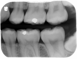
bitewing dental xray
- Periapical X-rays show the entire tooth, from the exposed crown to the end of the root and the bones that support the tooth. These are used to find dental problems below the gums such as impacted teeth, abscesses, cysts, tumors, and bone changes linked to some diseases.

periapical x-ray
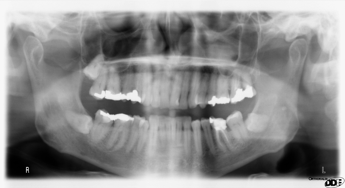
- Panoramic X-rays like that above show a broad view of the jaws, teeth, sinuses, nasal area, and temporomandibular (jaw) joints. These X-rays do not find cavities but do show problems such as impacted teeth, bone abnormalities, cysts, solid growths (tumors), infections, and fractures.
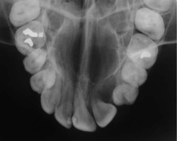
occlusal x-ray
Dangers Dental X rays
|
The amount of radiation used in dental X-rays is low, but there is always a slight risk of damage to cells or tissue from being exposed to any radiation, including the low levels of radiation used for this test. The risk of damage from the X-rays is usually very low compared with the potential benefits.
Pregnant women may not want to have routine dental X-rays taken until after they give birth. Although there is no proof that a routine dental X-ray could harm a developing baby (foetus). Dentists usually suggest you wait to have your X-rays until after the baby is born. Delaying the X-ray for a few months will not result in further harm to teeth in most cases. There are times when the severity of the dental problem requires an X-ray to deal with an urgent concern. Why might you not be able to have a dental x-ray taken?
|
*BUT WHY DOES THE DENTIST RUN OUT OF THE ROOM WHEN THEY TAKE THE X-RAY?*
The dental team might take hundreds of X-rays every week. Staff limit the amount of radiation they receive by moving away from the X-ray beam. However, the risk to patients from one or two routine X-rays is tiny. Staff check how much radiation they are exposed to by wearing a small badge during working hours. This is sent off to be analysed at regular intervals.
Advances in dentistry over the years have lead to the low radiation levels emitted by dental X-rays. Some of the improvements are new digital X-ray machines that limit the radiation beam to the small area being X-rayed,
By law in the UK, annual checks on all aspects and safety, as well as annual updating of training for all staff using dental x ray equipment is mandatory so rest assured you are in safe hands |
Who's Property are the X rays?
X-rays are an essential part of your health records and must be kept with your personal dental file. As dental records work differently to normal health records, your dentist must keep your dental records for at least two years from the date of your last course of treatment. You are entitled to copies of your records and X-rays under the Access to Health Records Act 1990. But you will have to pay for these copies. In most cases your X-rays and records will not be needed by your new dentist.
My Dentist wants to do a CT scan, WHY?
CT (computed tomography) is an imaging technique used in cases where normal 2d x-rays do not give enough information for effective, safe treatment planning and are increasingly used in implant cases in the UK.
Dental surgeries with this facility use cone beam CT which is a type of CT scanner with a much lower radiation dose compared to a normal medical CT scanner. The scanner goes around your head often in a similar way to the large x-ray at the top of this page it takes a lot of 2d slice images and converts them into a 3d image
As you can see below, the information means widths of bone can be measured accurately among other things and from these quite often templates can be made so as implants are placed in exact positions!
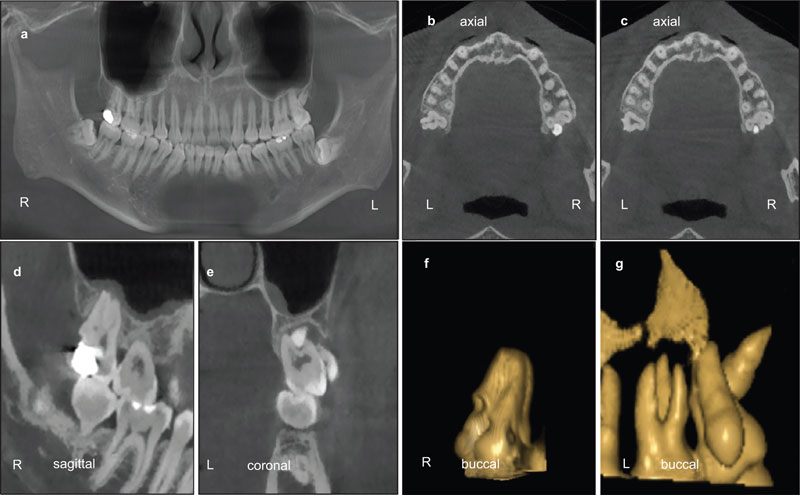
CT scan of jaws showing different views
SO HOW OFTEN DO I NEED X-RAYS/RADIOGRAPHS AT THE DENTIST?
|
Adults at low risk of tooth decay
|
Children at low risk of tooth decay
|
 RSS Feed